

Currently, specialists in the field of medicine have a lot of methods by which you can make an accurate diagnosis. One of them is an MRI or magnetic resonance therapy. Compared to the X -ray, this method allows you to visualize the organs in several planes and get clear pictures at once. MRI of the abdominal organs allows you to conduct an examination in order to obtain accurate results.
Content
- MRI of the abdominal cavity - about the method
- Indications or who needs to make an MRI of the abdominal cavity?
- What organs are checked with an MRI of the abdominal cavity
- Are there any contraindications to the abdominal MRI?
- Preparation for MRI abdominal cavity
- How is the abdominal MRI.
- What shows an MRI of the abdominal cavity and retroperitoneal space
- Which is better than CT or MRI of the abdominal cavity
- Ultrasound or MRI of the abdominal cavity, which is better
An examination is carried out on a special apparatus, which allows you to obtain contrasting and clear sections of organs (including the abdominal cavity) and facilitate the diagnosis without harm to humans. When compared with the old method - an X -ray, then this species allows you to make a diagnosis as accurately as possible.
MRI of the abdominal cavity - about the method
MRI allows you to find out more information about organs in comparison with other methods: X -ray pictures, ultrasound and laboratory indicators. When deciphering MRI of the abdominal cavity, specialists take into account the results of previous examinations.
The price of an MRI of the abdominal cavity is about 4-8 thousand rubles for the study of one or more organs. For example, the cost of an MRI of the abdominal cavity with contrast costs from 4 to 7-7.5 thousand rubles.
Indications or who needs to make an MRI of the abdominal cavity?
If we consider the standard method, that is, an MRI without contrasting is completely safe for a person. Even if the patient needs urgent diagnosis during exacerbation, MRI is safe. The magnetic field does not cause complications and does not have side effects, which cannot be said about the X -ray. That is why MRI is allowed to make people of any age, as well as children, especially if you need to make an accurate diagnosis.
Who recommended the examination? If a person’s diseases of the abdominal organs are diagnosed, treatment is carried out or surgery is necessary - the examination allows the diagnosis as accurately as possible.
In the event that pain in the abdomen is worried, and standard research methods do not show the causes (ultrasound and x -ray), MRI is also recommended.
Indications:
- pancreatitis;
- jaundice;
- if the abdominal cavity has a clusting accumulation;
- with pathological changes in internal organs. For example, necrosis, fibrosis, cirrhosis and liver dystrophy;
- if the liver and spleen increased in size;
- in case of damage to internal organs;
- anomaly of the development of the abdominal organs;
- neoplasms on internal organs: cysts, lipomas, adenomas; Oncological neoplasms of the primary nature and metastatic.
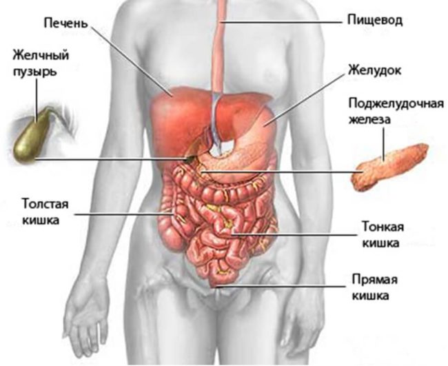
What organs are checked with an MRI of the abdominal cavity
Abdominal systems and organs:
- pancreas;
- liver and ducts with gallbladder;
- spleen;
- small and large intestines;
- stomach;
- lymph nodes and vessels, as well as soft tissues of the walls of the abdomen and abdominal cavity.
The organs of the retroperitoneal space:
- paranephral fiber;
- adrenal glands;
- kids.
MRI of the abdominal cavity: What is included:
- The study of the biliary tract and the bubble allow: to evaluate the condition of these organs, ducts, diagnose the presence of stones, detect neoplasms.
- Examination of urinary tract and kidneys in order to identify tumors, causes of anuria and hematuria, in order to detect stones in the kidneys, their number, location and size, diagnosis of inflammatory processes (for example, pyelonephritis), renal colic, and the state of internal organs during injury.
- A study of the pancreas, spleen and liver in order to detect foci of inflammation, cysts, tumors (good and malignant), metastasis, examination of metabolic processes in organs, diagnosis of organs for damage and injury.
- The stomach: diagnosis of lymph nodes and neoplasms, the body in order to make an accurate diagnosis (for example, malignant formations and their degree).
- Vessels: in order to identify anomalies of development and structure, deviations.
- Lymphatic system: in order to detect inflammatory processes, metastasis (localization, size), diagnosis.
Magnetic resonance therapy is a modern type of examination of the abdominal organs in order to make an accurate diagnosis. For example, after obtaining the results of ultrasound, x -ray and laboratory tests, the doctor doubts the size of neoplasms, the focus of inflammation or anomaly of the structure of the organs. For this purpose, an MRI is prescribed in order to make the diagnosis as accurately as possible and, for example, to understand what nature of the neoplasm (malignant, benign). Now you know which abdominal organs can be examined using MRI.
Are there any contraindications to the abdominal MRI?
Despite the fact that MRI is a relatively safe type of examination, but there are restrictions. The procedure is not carried out if there are metal elements in the human body: pacemakers, insulin pumps, vascular clips, hearing aids with metal elements, prostheses, pins, etc., that is, everything that can be attracted by the magnet.
If the patient did not notify the doctor of the presence of metal objects in his body, this can negatively affect the work of the tomograph. Under the influence of an electromagnetic wave, the devices are heated and can break down.
These were absolute contraindications, but there are also relative ones, which are also undesirable to conceal, this is:
- fear of closed space;
- anxiety, panic attacks;
- convulsive seizures (epilepsy);
- hemodialysis;
- strong pains in which a person cannot lie motionless in one position for a long time.
In addition, it is worth considering that:
- if it is necessary to conduct a study using a contrasting liquid, then MRI cannot be carried out during breastfeeding and pregnancy;
- the weight of the patient is more than 130 kg - in this case, conduct a study or not, the attending physician decides;
- kidney diseases (using contrasting liquid);
- individual intolerance - if a contrast medium is introduced.
The doctor must notify the patient about all the restrictions before the study. If a person is afraid of a closed space, afraid of monotonous sounds and noise, it is necessary to inform his attending physician.
Preparation for MRI abdominal cavity
The study of the abdominal MRI is a simple procedure that does not harm the health of the patient. But in order to get the most accurate results, it is necessary to carefully prepare.
Rules for preparing for MRI:
- put comfortable clothes that will not interfere, crush and constrain movements. There should not be any metal elements in this clothing, that is, you can not wear a sweater on a snake, clothes with metal inserts, etc.;
- 24 hours before the set date of the examination, you can not drink carbonated water and drinks. It is impossible: tea and coffee, fruits and vegetables, brown bread and all other products that can cause gas formation;
- if the liver, pancreas and spleen are examined, carbohydrates are excluded;
- general rules of preparation: the last meal 8 hours before the examination;
- drink any liquid, including purified water - the last time you can drink 5 hours before the examination;
- if a diet is observed, but the patient has increased gas formation, for the purpose of prevention, the attending physician can prescribe special drugs: activated carbon or escumizan.
- 30 minutes before the start of the examination, it is necessary to take an antispasmodic.
If the case is emergency, the patient is allowed to undergo an examination without preparation, but there is no guarantee that the results of the examination can be as accurate as possible.
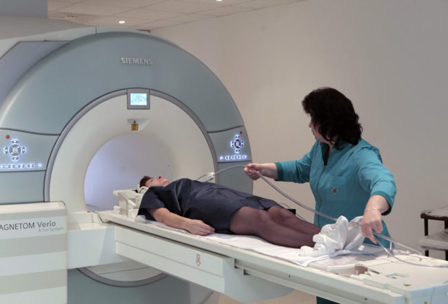
How is the abdominal MRI.
MRI is not a quick method of examination, it is necessary to lie without moving within half an hour or even more, it all depends on which area is examined.
During the examination, a doctor may offer to put on headphones and give a sensor with a button for communication. During the examination, the doctor will speak when you need to hold your breath. Breathing holding the maximum of 20 seconds.
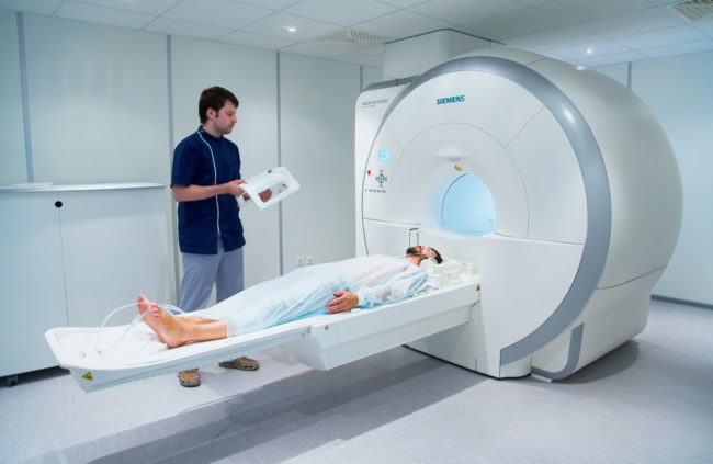
The patient may be offered to fix the limbs so as not to control his body during the examination. If necessary, the contrasting substance for the accuracy of the examination is intravenously.
The patient should be ready for the fact that for half an hour he will be in a closed capsule, but there is lighting and ventilation inside, and there will also be the opportunity to communicate with doctors.
What shows an MRI of the abdominal cavity and retroperitoneal space
Magnetic resonance therapy allows you to identify deviations in the work of the abdominal cavity, if there are suspicions of tumors and metastases, in order to exclude and identify severe liver diseases and pancreas, with any injuries (long and fresh), if there are suspicions of the development of internal organs. And suspicions of bleeding. MRI is also prescribed before planned surgery, if standard examinations were not enough.
When examining the organs of retroperitoneal space, it is possible to identify deviations in the work of the kidneys, adrenal glands, ureters in order to clarify the diagnosis, identify neoplasms (cysts, tumors, metastases), developmental abnormalities, and control the treatment process in the fight against cancer.
Which is better than CT or MRI of the abdominal cavity
CT or computed tomography is a study of the body using x -rays. But this is not an ordinary x -ray, when the rays pass through the patient’s body, focusing on a film, this is a modern examination method that allows you to get a voluminous image.
Briefly, the study is carried out as follows: the patient lays on the couch, above which the source of X -rays of the ring -shaped circuit is installed. As a result of the activation of the device, pictures from different angles and points are taken, which are then processed on the computer and a modeled image of the body under study is obtained.
MRI examination is the same principle, only the image of the organ will be three -dimensional. It is impossible to say exactly which examination is impossible, because each method has its own characteristics, advantages and disadvantages.
Differences and possibilities of methods:
- Computed tomography allows you to detect damage to teeth, bones, joints, diagnose injuries and diseases of the spine (hernia, scoliosis, etc.), detect deviations in the brain, examine the chest organs in order to clarify the supply of diagnosis, examine the thyroid gland, blood vessels, organs genitourinary system. We can say that CT is a universal diagnostic method, but in order to make the most accurate diagnosis, the examination is carried out using a contrasting substance.
- Magnetic resonance therapy is more suitable for the study of blood vessels, joints and soft tissues, organs of the abdominal cavity.
In short, we propose to get acquainted with the readings and contraindications of both methods:
- the computed tomography is directed with fractures and complex injuries, in order to detect bleeding of internal organs, to examine the work of the stomach and lungs;
- on the MRI is directed if you need to diagnose inflammatory processes, tumor formations, diagnose the state of the nervous system.
The contraindications of MRI were said above, and this method is prohibited from conducting in such cases:
- during pregnancy;
- breastfeeding;
- with improper work of the kidneys;
- diabetes mellitus;
- thyroid diseases;
- if a person weighs 150 kg and more;
- if the patient has an uncontrolled condition or he is excited.
CT examination lasts no more than 15 minutes, and MRI - a maximum of 40. If the examination is necessary for the child, then with CT parents can be near. With an MRI in the capsule, there is only one person. Inside the camera, the patient can be scared of clicks and noise, which makes the device, so the doctor offers to put on headphones and shows a button for communication with a specialist.
On safety: CT is carried out using contrasting fluid, and this is the minimum dose of irradiation.
Ultrasound or MRI of the abdominal cavity, which is better
An ultrasound study is the most common type of examination of the abdominal organs. It takes no more than a quarter of an hour in time, in some cases it requires special training. Ultrasound allows you to identify violations in the work of internal organs, identify neoplasms and see internal bleeding.
There is an ultrasound for:
- gynecological and obstetric examination;
- in order to inspect the state of internal organs: kidneys, stomach, liver, thyroid gland, etc.
Considering the question of which examination is better, it is worthwhile to understand that an ultrasound is a quick way, and an MRI - when it is necessary to conduct a more extensive examination in order to clarify the diagnosis.

If there is a choice of what to make an MRI or ultrasound, keep in mind that in the second case the responsibility completely falls on a specialist and even the most qualified employee can make an error and make an inaccurate diagnosis. For MRI, the results are processed on the computer, errors and inaccuracies are excluded.


Comments
a couple of years ago, there was no side of metrogils from the same problem, there were no side effects ...
I’m not a fan of peeling at all, it saves from acne of metrogil, it also smoothes it ...
Great article! ...
I take the second course of the Capsules Climafite 911. The tides went very quickly. It became calmer, irritability went away and I sleep well ...
i also noticed - it is worth nervous, everything immediately affects the face. Therefore, I try to avoid conflicts and unpleasant people. Of the creams, I like Miaflow from wrinkles - smoothes not only small wrinkles ...